

Translate this page into:
Uncommon Case of Supernumerary Teeth and their Management: A Rare Clinical Case Report

* Corresponding author: Dr M M Dayakar, Prof and HOD, Department of Periodontology, K.V.G Dental college & Hospital, Kurunjibhag, Sullia, Karnataka, India. mmdayakar@yahoo.com
-
Received: ,
Accepted: ,
How to cite this article: Dayakar MM, Dayakar A, Hemanth P. Uncommon Case of Supernumerary Teeth and their Management: A Rare Clinical Case Report. Dent J Indira Gandhi Int Med Sci. 2024;3:112-4. doi: 10.25259/DJIGIMS_18_2024
Abstract
Supernumerary teeth are often classified into four groups: conical, supplemental, tuberculate, and odontomas. Supernumerary teeth of the tuberculate kind typically have many cusps or tubercles on the tooth crown. This sporadic eruption into the oral cavity may lead to the maxillary central incisors impaction. The purpose of this article is to discuss surgical management, various problems associated with dental overcrowding, and the need to detect excess teeth early on, as they can cause space loss and impaction of the incisors.
Keywords
Supernumerary
Diastema
Malocclusion
Tuberculate

INTRODUCTION
Typically, the central incisors are the ones that erupt into the oral cavity first.[1] However, in this uncommon instance, the child’s permanent teeth had all erupted into the oral cavity, with the exception of the maxillary right central incisor. There are various complications associated with the presence of such supernumerary teeth in the oral cavity.[2] Usually found in the maxilla close to the midline, a mesiodens is the most common type of supernumerary tooth. It could result in crowding, ectopic growth, mid-line diastema, incisor rotation, ectopic eruption, periodontal disease, caries, or impaction of the incisor tooth or teeth. On rare occasions, it may lead to the development of a dentigerous cyst or the resorption of the neighboring tooth’s root.[3,4] These extra teeth may erupt into the maxillary sinus or nasal cavity, or they may be impacted and/or erupted.[4,5] In other situations, they might not result in any pathological conditions, but they still need to be taken out to relocate teeth in orthodontics, graft bone in cases of cleft lip and palate, or place implants in the jaw.[6,7]
Some authors advise exposing the impacted permanent tooth and extracting the supernumerary tooth at the same time if the supernumerary teeth caused the incisors next to it to become impacted.[8] The impacted incisor is exposed when the supernumerary tooth is removed, and in about 78% of instances[9] and 30% of cases, the incisor spontaneously erupts within 16 months. Orthodontic traction is necessary in certain instances. Complex surgery and orthodontic therapy are necessary only when the impacted tooth is deep in the bone, badly displaced, the arch is too small, or the affected tooth shows full root formation. If a supernumerary tooth in primary dentition does not cause any significant issues, its presence should be informed to the patient or their guardians, and they will decide to keep it or get it extracted.
Case Report
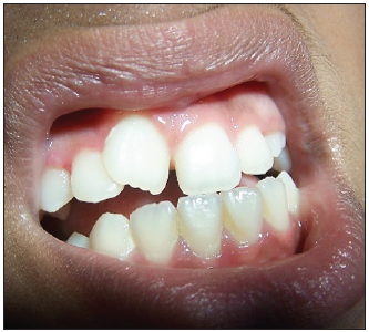
Deekshit, a 12-year-old boy, was complaining of missing upper front teeth [Figure 1]. Upon clinical examination, it was discovered that there was significant space loss in the incisor region and that the upper central incisor was absent from the dental arch due to impaction. Impaction of the maxillary right central incisor was visible in the intra-oral periapical radiograph [Figure 2], along with the uncommon presence of a supernumerary tooth on the crown of the already impacted maxillary central incisor. Parents were informed of the problem and gave their consent for surgery. Routine blood and urine tests were performed before surgical procedures, and the findings were within normal ranges. Adequate local anesthesia was given, with a 15c scalpel blade, and a full-thickness labial mucoperiosteal flap was raised. Impacted supernumerary teeth were extracted using a periosteal elevator [Figure 3a-b]. After thorough irrigation, the flap was sutured, and seven days later, the patient was recalled for suture removal and follow-up. Post-operative instructions suggested using an antiseptic mouthwash, prophylactic antibiotic coverage, and maintaining proper oral hygiene. After three months, the affected maxillary right central incisor erupted successfully, but the presence of less space led to malalignment of the tooth [Figure 4].
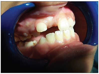
- Pre-operative photograph showing the impacted of the maxillary right central incisor.
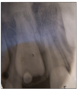
- Intraoral Periapical Radiograph (IOPAR) showing impacted the maxillary right central incisor with 1 impacted supernumerary tooth on it.
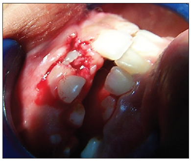
- Extraction of supernumerary teeth.
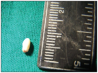
- Presence of tuberculate and underdeveloped root.
- Erupted maxillary right central incisor but crowded after 3 months.
DISCUSSION
Prior to performing any surgery, a comprehensive clinical and radiographic assessment of the affected supernumerary tooth should be completed. The radiographs consist of a maxillary occlusal film, an intra-oral periapical radiograph (ideally using a buccal object approach), and a panoramic radiograph. Precise radiographs are essential for ascertaining the location of the affected tooth, its relationship to neighboring teeth, and the state of the roots nearby. To identify supernumerary teeth in three dimensions and to identify neighboring tooth root resorption, computed tomography (CT) is more precise; nevertheless, from the perspective of the Indian economy, it will be expensive. Any kid who is suspected of having an impacted supernumerary tooth or teeth might have a delayed eruption, or impaction of teeth or other teeth and should receive an early diagnosis and appropriate interceptive management. This will lessen the possibility of malocclusions developing or the production of dentigerous cysts [3,4] or resorption of neighboring teeth roots, which will allow permanent teeth to erupt spontaneously into their location.
On the other hand, if we delay the preventive and interceptive treatment due to an impacted supernumerary tooth that caused impaction, it will lead to losing tooth space for the eruption of permanent teeth and could easily result in space loss in the dental arch and necessitate a complicated treatment strategy later.[10] Supernumerary tooth formation is believed to have a multifactorial etiology. Several authors have documented the genetic influence on its development, yet it deviates from the straightforward Mendelian pattern of inheritance. Many syndromes like Gardner syndrome, Down syndrome, cleidocranial dysplasia, and cleft lip and palate, which are strongly associated with inheritance, are typically linked to multiple extra teeth. Certain authors suggest that many supernumerary teeth are caused due of the hyperactivity of the dental lamina. The authors also proposed that a third tooth bud next to the permanent tooth in the dental lamina might develop into a supernumerary tooth. According to its original theory, tooth buds are separated into two components that are equal or not, forming two teeth that may grow equal in size or dysmorphic in size and shape. This concept is known as the “dichotomy theory.” Animal experiment where split tooth germs have been grown in vitro lends credence to this theory. Whether a supernumerary tooth is erupted or impacted in the dental arch in permanent dentition most often creates an un-aesthetic appearance for the child, which concerns the parents. According to this case report, children’s appearance can be significantly enhanced by modest orthodontic treatment and the spontaneous eruption of impacted teeth following surgical extraction of supernumerary teeth.
CONCLUSION
In developing children, supernumerary teeth can lead to a variety of malocclusions and other issues. The dentist must diagnose the disease early and accurately, provide early detection, and administer preventative and interceptive care. This will lessen the worry that parents and children with the disorder experience while also improving the child’s appearance.
Ethical approval
Institutional Review Board approval is not required.
Declaration of patient consent
The authors certify that they have obtained all appropriate patient consent.
Financial support and sponsorship
Nil.
Conflicts of interest
There are no conflicts of interest.
Use of artificial intelligence (AI)-assisted technology for manuscript preparation
The authors confirm that they have used Artificial Intelligence (AI)-Assisted Technology for assisting in the writing or editing of the manuscript or image creations.
REFERENCES
- The unerupted incisors. A study of post-operative eruptive history of incisors delayed in their eruption by supernumerary teeth. Dent Pract Dent Rec. 1967;17:332-341.
- [PubMed] [Google Scholar]
- Shafer’s textbook of oral pathology (5th ed). Netherlands, Europe: Elsevier; 2006. p. :1-1280.
- Dentigerous cyst due to mesiodens: report of two cases. J Ir Dent Assoc. 1989;35:117-118.
- [PubMed] [Google Scholar]
- A rare cause of nasolacrimal obstruction: Dentigerous cyst in the maxillary sinus. Indian J Ophthalmol. 2009;57:465-7.
- [CrossRef] [PubMed] [Google Scholar]
- Orthodontic-Principle and Practice.
- Pediatric oral and maxillofacial surgery.
- The effect of variation in tooth morphology and position on eruption. Dent Pract Dent Res. 1971;22:95-108.
- [Google Scholar]
- Supernumerary teeth and their management in children. Int J Oral Health Med Res. 2016;3:32-7.
- [Google Scholar]
- Supernumerary teeth causing delayed eruption, a retrospective study. Br J Orthod. 1992;19:41-6.
- [CrossRef] [PubMed] [Google Scholar]
- The distribution of anomalies of primary anterior teeth and their effect on the permanent successors. Dent Clin North Am. 1984;28:69-80.
- [CrossRef] [PubMed] [Google Scholar]


