

Translate this page into:
Salvage Through Hemisection – A Case Report


Corresponding author: Dr. Sulaima Mehar, Department of Conservative Dentistry and Endodontics, Mithila Minority Dental College and Hospital, Darbhanga, Bihar, India. meharkhan357@gmail.com
-
Received: ,
Accepted: ,
How to cite this article: Mehar S, Sengupta R, Kumar A, Jethwani J. Salvage Through Hemisection – A Case Report. Dent J Indira Gandhi Inst Med Sci. 2023;2:130–3. doi: 10.25259/DJIGIMS_11_2023
Abstract
Advances in dentistry and the increasing desire of patients to preserve their teeth have led to the treatment of teeth that would previously have been removed. The first molars of the lower jaw are the teeth that are most frequently extracted due to caries and periodontal disease. These teeth are the most important position for occlusion and also have a large pericemental area. Therefore, for any defect at the root, whether mesial or distal, extraction is the most commonly planned treatment. Under certain conditions, only the diseased part of the tooth can be extracted after endodontic treatment. A modified fixed partial denture is fabricated to splint the remaining part of the tooth to the adjacent teeth. Although this procedure is demanding, it is easy to perform and can be successfully maintained.
Keywords
Furcation defects
Root resection
Caries

INTRODUCTION
With the evolution of modern practices in dentistry, the opportunity to maintain a functional dentition for a lifetime has become much easier. Not only is it a satisfying approach for the patient in terms of expenses in comparison to replacement with a dental implant or a chance of treatment failure, but above all, preserving something which is absolutely natural is a good thing in every aspect.[1]
A grossly decayed molar substitute as a terminal support is generally inappropriate for restoration. In such patients, providing a removable partial denture or fixed prosthesis like a dental implant is desired to substitute the missing tooth.[2] Nevertheless, in cases where the decay is confined to just one of the roots, a hemisection can be a good alternative. But before starting with any procedure, a case selection must first be made after doing endodontic, periodontal, and prosthodontic assessments of the tooth.
From a periodontal point of view, if the tooth has extensive bone loss confined to one of the roots or contribution of Class 3 furcation defect that has the probability of becoming stable after the removal of one root, then that case can go for hemisection.[3] Also, this process is allowed if the patient has a negligent attitude towards maintaining oral hygiene or is incapable of doing so. Periodontal defects like dehiscence are an alternative indication for the removal of one root.[4] From a restorative point of view, hemisection is favorable in cases where there are chances that abutment failure will not occur if one root is removed, and the other is absolutely healthy in serving as an abutment for fixed prosthesis.[5] Additional suggestions can be vertical root fracture of one root, any destructive process like caries, external root resorption, trauma, etc., confined to just one root of multi-rooted teeth. Contraindications are fused root, compromised second root and the remaining root inoperable for obligatory root canal treatment.[6,7]
Hemisection represents a form of conservative dentistry that aims to retain as much of the original tooth structure as possible. It refers to the division of the mandibular molar in two halves monitored by the elimination of the compromised root along with its coronal part.[8-10]
CASE REPORT
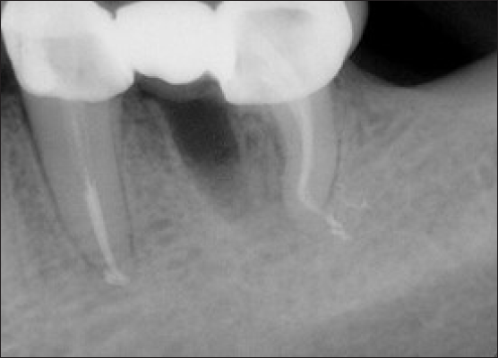
A 35-year old male came to the Department of Conservative Dentistry and Endodontics with complaints of food lodgement and pain in the lower left first molar tooth. On intraoral examination, the tooth was grossly decayed, restored on the mesio-proximal side, there was tenderness on percussion, mobility was within physiological limits and with sound periodontium [Figure 1]. On radiographic examination, disto-proximal caries were seen in 35, dislodged restoration was seen in 36, with caries extending up to the mesial root of 36. The bony support of the tooth was intact [Figure 2].

- Pre-operative photograph of the molar
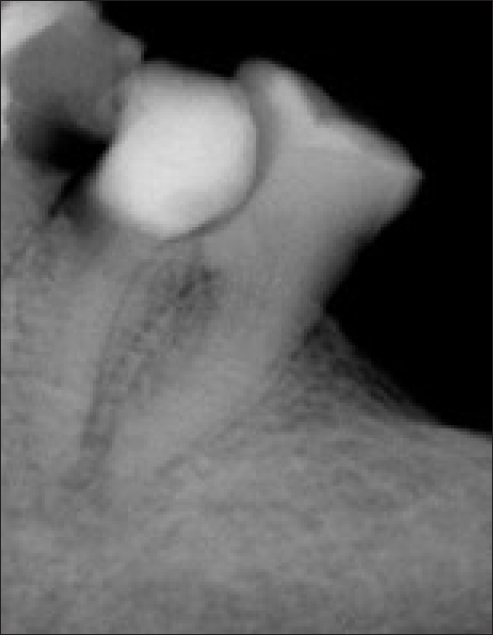
- Pre-operative radiograph with regards to 36
Hemisection of the mesial root of 36 was decided Post-endodontic treatment. Root canal treatment was done in 35 and 36. After doing the access cavity with EndoZ bur (Dentsply, Sirona), hand instrumentation till 15# k file was done. Neoendo S rotary file was used in sequence till 25 4% in mesio-buccal root of 36, and the disto-buccal root was prepared with 25 6% [Figure 3]. Mandibular premolar was prepared till 35 4%. Sodium hypochlorite of 5.25% concentration was used for intermittent irrigation using a side vented needle, and 17% ethylene diamine tetra acetic acid (EDTA) was used as a final rinse solution; AH plus sealer was used for obturation. Glass fiber post was placed in 35 [Figure 4].
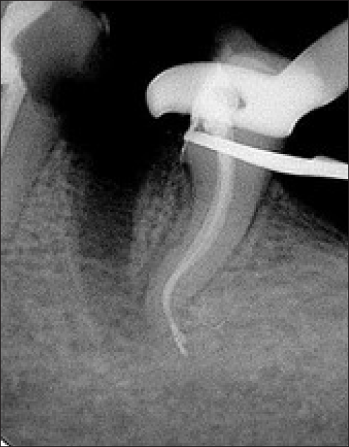
- Radiograph after removal of mesial root with regards to 36
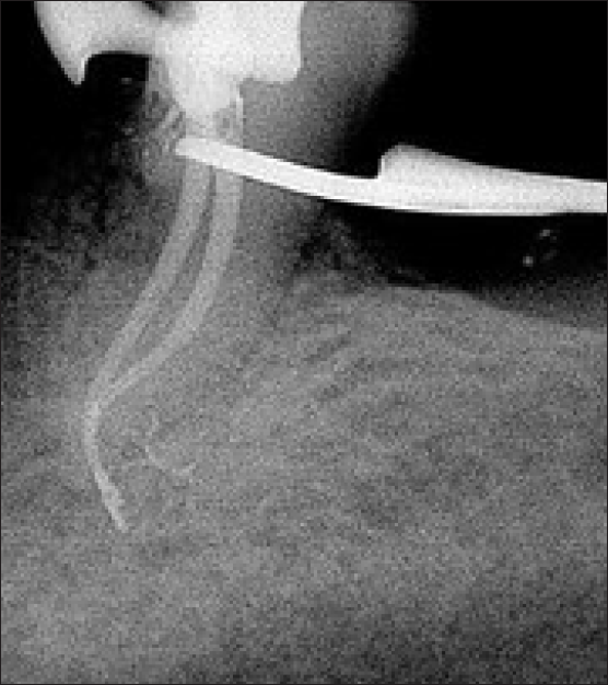
- Obturation radiograph of distal root with regards to 36
With the help of a tapered fissure bur, an incision was made vertically in a bucco-lingual direction, taking care not to traumatize the bone and the adjacent tooth. The socket was cleaned and irrigated with normal saline, and root planing of the distal root of 36 was done.
Restorative plan
Post and core were done in 35. A three-unit fixed partial denture was given, including the distal root of 36 [Figure 5].
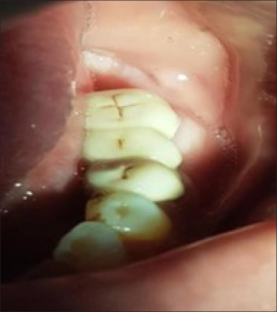
- Post-operative photograph after luting of prosthesis
- Post-operative radiograph
DISCUSSION
According to Buhler, hemisection is a steadfast alternative to extraction in the mandibular molar, and root canal treatment must be completed prior to this procedure to ensure a long-term treatment outcome with positive results. In the case of endodontic breakdown, hemisection should be considered a failure.[3]
Three factors important for considering a tooth for hemisection are: 1) the root has to be divergent, 2) the furcation opening has to be closer to cemento enamel junction (CEJ) and 3) there should be proper bony contour; otherwise, odontoplasty has to be done.
Buhler et al. found a 32% failure rate of hemisection at 10 years.[11] The primary etiology behind it is endodontics pathology and root fracture. In a follow-up of 3–10 years, Blomlof et al. discovered the same failure rate. In a study by Park et al., hemisection is a bit dependable option for molars with doubtful prognosis as it can save the teeth from significant bone loss for a long period of time if the patient’s oral hygiene is good.[12] In a study by Shafique et al., hemisection was found to be a valuable alternative in mandibular molars where one root is compromised by decay, and the other one is healthy enough to act as an abutment. Carnivale, in his study on the long-term effects of root resection therapy, concluded that periodontal problems like furcation defects can be easily managed by hemisection.[13]
CONCLUSION
The procedure of hemisection requires precise knowledge and good professional skills. Careful case selection is a way to long-term clinical success in the future.
Declaration of patient’s consent
The authors certify that they have obtained all appropriate patient consent.
Financial support and sponsorship
Nil.
Conflicts of interest
There are no conflicts of interest.
Use of Artificial Intelligence (AI)-Assisted Technology for manuscript preparation
The authors confirm that there was no use of Artificial Intelligence (AI)-assisted technology for assisting in the writing or editing of the manuscript and no images were manipulated using AI.
REFERENCES
- Endodontic Therapy (5th Edition).
- Survival rates of hemisected teeth: An attempt to compare them with survival rates of alloplastic implants. Int J Periodontics Restorative Dent. 1994;14:536-43.
- [PubMed] [Google Scholar]
- Vital hemisection of a mandibular second molar:A case report. J Am Dent Assoc. 1981;102:503-6.
- [CrossRef] [PubMed] [Google Scholar]
- Hemisection-technique and restoration. Dental Clinics of North America. 1974;18:15-44.
- [PubMed] [Google Scholar]
- Furcation involvements: Therapeutic considerations. Compend Contin Edu Dent. 1998;19:1236-40, 1242, 1244.
- [PubMed] [Google Scholar]
- Traumatic hemisection and restoration of a maxillary first premolar: A case report. General Dentistry. 2003;51:340-2.
- [PubMed] [Google Scholar]
- Hemisection as an alternative treatment for decayed multirooted terminal abutment: A case report. J Canadian Dent Asso. 2009;75:387-90.
- [PubMed] [Google Scholar]
- Evaluation of root-resected teeth. Results after 10 years. J Periodontol. 1988;59:805-10.
- [CrossRef] [PubMed] [Google Scholar]
- Hemisection of teeth with questionable prognosis. Report of a case with seven-year results. J Int Acad Periodontol. 2009;11:214-9.
- [PubMed] [Google Scholar]
- Management of furcation involvement. Periodontol 2000. 1995;9:69-89.
- [CrossRef] [PubMed] [Google Scholar]


