

Translate this page into:
Management of Pyogenic Granuloma in the Mandibular Anterior Region Using Diode Laser

*Corresponding author: Dr. Gayathri Priyadharshini Elangovan, Department of Periodontology, Vivekanandha Dental College For Women, Ellayampalayam, Tiruchengode, Tamil Nadu, India. gayathriaelangovan@gmail.com
-
Received: ,
Accepted: ,
How to cite this article: Elangovan GP, Periyasamy IK. Management of Pyogenic Granuloma in the Mandibular Anterior Region Using Diode Laser. Dent J Indira Gandhi Int Med Sci. 2025;4:39-41. doi: 10.25259/DJIGIMS_38_2024
Abstract
Pyogenic granuloma is the most common tumor-like growth that occurs in the oral cavity. It is expressed as reactive hyperplasia of the gingival connective tissue in response to etiological factors such as local irritation, trauma, and hormonal factors. Other factors accused of causing pyogenic granuloma include foreign materials, hypertension, and poor oral hygiene. The pyogenic granuloma most commonly occurs in the gingiva. Pyogenic granuloma in the mandibular anterior region is associated with poor aesthetics and discomfort as it interferes with mastication. The purpose of this case report is to evaluate the management of the pyogenic granuloma diagnosed in a 25 year old male patient using a diode laser.
Keywords
Pyogenic granuloma
Pregnancy tumor
Gingivectomy
Management of Pyogenic granuloma
LASER excision

INTRODUCTION
The first reported case of pyogenic granuloma was described in the year 1884 by Hullien[1], and the term pyogenic granuloma was coined in 1904 by Hartzell. Pyogenic granuloma is a mucocutaneous lesion that is benign and nonneoplastic in nature, and occurs in the skin and mucous membranes.[2] It occurs mostly in the second and third decades of life.[3] The pyogenic granuloma is most commonly found in the gingiva, but it sometimes occurs on the buccal mucosa, lip, palate, and tongue.[3] Pyogenic granuloma appears more frequently in the maxillary anterior region.[4] It occurs more frequently in females than in males, especially during pregnancy.[5] It is generally believed that female sex hormones play an important role in the pathogenesis of the pyogenic granuloma.[5] It appears clinically as a soft, smooth, lobulated tumor-like mass with or without the pedicle.[6]
The etiological factors for pyogenic granuloma are responses to local irritation, trauma, and hormonal factors. Other factors accused of causing pyogenic granuloma include foreign materials, hypertension, and poor oral hygiene.[7] The purpose of this case report is to present a pyogenic granuloma found in the mandibular anterior region of a 25-year-old male, interfering with mastication and its management using a diode laser.
CASE REPORT
A 25-year-old male patient visited our department with a chief complaint of gingival growth in the lower front tooth region for the past two months. It wass associated with gingival bleeding on provocation. She reported a gradual increase in the size of gingival growth, which led to gingival bleeding and discomfort while eating and brushing, as the gingival overgrowth reached the occlusion and interfered with mastication. No extra-oral finding were detected in the patient and he was systemically healthy.
On intra-oral clinical examination, a well-defined, pedunculated, gingival overgrowth with inflammation was seen in the interdental papilla between 31 and 32. The swelling appeared reddish pink in color, ovoid in shape, and soft in consistency with a small erythematous papule. The overgrowth had a pedunculated base which bled on slight provocation, and there were no signs of tooth mobility in the 31st and 32nd region. [Figure 1]. An intra-oral periapical radiograph was taken for the lower anterior region and no signs of boneloss were seen. Routine hematologic tests were done for the patient and the report revealed that the results were within the normal range. With the above findings, we arrived at the provisional diagnosis of pyogenic granuloma.
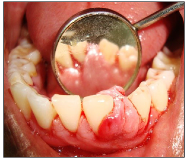
- Preoperative.
The treatment schedule was planned. During the first session, supragingival scaling was done, followed by irrigation of the 31st and 32nd regions using chlorhexidine. Surgical therapy was scheduled for after a week, and post-operative instructions were given to the patient. During surgical therapy, the treatment plan for this lesion was wide surgical resection of this lesion from its periphery using light amplification by stimulated emission of radiation (LASER). One week after the first session, the patient was recalled, and an excision of the lesion and gingivoplasty was done in relation to the 31st and 32nd regions under the diode laser in continuous mode [Figure 2]. The excised tissue was sent for histopathological analysis. Hemostasis was achieved and irrigation using saline was done in relation to the 31st and 32nd regions [Figure 3].

- Excised tissue.

- Immediate postoperative.
DISCUSSION
Pyogenic granuloma most commonly occurs as an inflammatory response due to any chronic irritation that occurs because of the accumulation of plaque and calculus due to poor oral hygiene, overhanging restorations, and trauma.[7] The second most important aetiology, with a prevalence of 5% to 8%, is the hormonal change that occurs during pregnancy.[5] In such a condition, pyogenic granuloma is also called a pregnancy tumor. Medications, such as cyclosporine, may also be involved in the aetiology of Pyogenic Granuloma.[8] Intra-orally, pyogenic granulomas have a greater predilection for gingival tissue, and the interdental papilla is the most common site. In our case, pyogenic granuloma was present in the interdental papilla of the 31st and 32nd regions. For the final diagnosis of pyogenic granuloma, the tissue has to be sent for biopsy.
The conventional treatment of pyogenic granuloma is the conservative surgical excision of the entire lesion. The reported recurrence rate for pyogenic granuloma is 16%. Other methods of lesion removal include cryosurgery, cauterization with silver nitrate, Nd: YAG, Diode, CO2 laser excision, and laser photocoagulation. Powell described using laser to excise lesions with the main advantage of no post-operative bleeding as compared to scalpel techniques.[9] Considering the advantages of laser in excision of the lesion, the patient was treated using diode laser.
Recently, Maffert et al introduced the use of a flash lamp pulsed dye laser for the excision of a mass of granulation tissue, such as pyogenic granuloma, which did not respond to the usual treatment methods.[10]
CONCLUSION
Pyogenic granulomas usually develop very rapidly, and the recurrence rate following treatment is very high . Hence, diode laser were used to completely remove them and prevent their recurrence. Other advantages of using diode lasers include no scar formation and less discomfort for the patients compared to other methods of excision.
Ethical approval
Institutional Review Board approval is not required.
Declaration of patient consent
The authors certify that they have obtained all appropriate patient consent.
Financial support and sponsorship
Nil
Conflicts of interest
There are no conflicts of interest.
Use of artificial intelligence (AI)-assisted technology for manuscript preparation
The authors confirm that there was no use of artificial intelligence (AI)-assisted technology for assisting in the writing or editing of the manuscript, and no images were manipulated using AI.
References
- Case of aneurism by anastomosis of the superior maxillare. Am J Dent Sci. 1844;4:160-162.
- [PubMed] [PubMed Central] [Google Scholar]
- Pyogenic granuloma subsequent to injury of a primary tooth. A case report. Int J Paediatr Dent. 2002;12:438-41.
- [CrossRef] [Google Scholar]
- Oral pyogenic granuloma: A case report and a comprehensive review. SRM J Res Dent Sci. 2015;6:257-60. Available from: http://dx.doi.org/10.4103/0976-433X.170284 [Last accessed on 2024 Nov 19]
- [CrossRef] [Google Scholar]
- Pyogenic granuloma--clinical features, incidence, histology, and result of treatment: Report of 242 cases. J Oral Surg. 1966;24:391-8.
- [PubMed] [Google Scholar]
- Clinical outline of oral pathology: diagnosis and treatment (3rd edn). Hamilton: BC Decker; 2002. p. :113-14.
- Pyogenic granuloma of the gingiva: A misnomer? - A case report and review of literature. J Indian Soc Periodontol. 2013;17:514-9.
- [CrossRef] [PubMed] [PubMed Central] [Google Scholar]
- Reactive hyperplasia of the oral cavity: A survey of 197 cases in Tabriz, Northwest Iran. J Dent Res Dent Clin Dent Prospects. 2010;4:87-9.
- [CrossRef] [PubMed] [PubMed Central] [Google Scholar]
- Treatment of pyogenic granuloma by shave excision and laser photocoagulation. Plast Reconstr Surg. 1999;104:1346-9.
- [CrossRef] [PubMed] [Google Scholar]
- Treatment of oral granulation tissue with the flashlamp pulsed dye laser. Dermatol Surg. 1998;24:845-8.
- [CrossRef] [PubMed] [Google Scholar]


