

Translate this page into:
Exploring Midline Asymmetry: A Comprehensive Study Among Adolescents in the North Bihar Population

* Corresponding author: Daya Shankar, Dentistry, Dental Medical Officer, Health Department, Bihar Government, Ex-Assistant Professor, Department of Dentistry, Patna Medical College & Hospital, Patna, Bihar, India. drdaya03@gmail.com
-
Received: ,
Accepted: ,
How to cite this article: Shankar D, Anjan R, Choudhary AK, Shankar R, Nanda M, Bharti G. Exploring Midline Asymmetry: A Comprehensive Study Among Adolescents in the North Bihar Population. Dent J Indira Gandhi Int Med Sci. 2024;3:9–12. doi: 10.25259/DJIGIMS_5_2024
Abstract
Objectives
In the contemporary era, achieving a sense of equilibrium is a paramount concern, and a visually pleasing smile is intricately linked to both professional success and social acceptance. Esthetics, with a specific focus on Oro-facial aesthetics, emerges as a primary motivation for individuals seeking dental treatment. Nevertheless, there exists an optimal and aesthetically pleasing range of dental midline deviation.
The current study aimed to ascertain the prevalence of dental midline shifting and the average extent of midline discrepancy among adolescents (aged 13-17) from North Bihar population.
Material & Methods
Two hundred subjects participated in the study, with 100 males and 100 females selected based on predetermined inclusion criteria. Intraoral frontal occlusion photographs were captured for each participant, and subsequent analysis was done utilizing Digimizer software. Statistical analysis was applied to the linear measurement data obtained through photographic evaluation.
Results
The study’s findings revealed a noteworthy proportion of subjects displaying deviations between upper and lower midlines and anterior teeth midlines, falling within the range of 0.03-3.80 mm.
Conclusion
The prevalence of these deviations was observed to be higher in males compared to females, and this difference was statistically significant.
Keywords
Esthetics
Asymmetry
Dental midline
Malocclusion
Occlusion

INTRODUCTION
One of the primary challenges frequently encountered by dentists and orthodontists is midline deviation, a condition where the dental midline, defined as the line bisecting the maxillary and mandibular dental arches within the midsagittal plane, does not coincide. While each dental arch possesses its own midline, noncoincidence of these lines results in midline deviation, a persistent issue that rarely corrects itself. Although historically viewed primarily as an esthetic concern, Jayalakshmi et al. 2013,[1] midline deviation holds significance beyond esthetics, indicating abnormal bilateral occlusion Levin. 1978.[2] Occlusion is recognized as a dynamic and functional relationship rather than a static condition, constantly influenced by various components of the masticatory system and subject to lifelong modifications Turp et al. 2008.[3] Understanding the intricate connection between occlusal functions and the evolving midlines is crucial for clinicians.Being cognizant, this study aims to assess and evaluate the incidence of midline asymmetry among adolescents from the North Bihar population.
AIM AND OBJECTIVE OF STUDY
Assessing the prevalence of dental midline shifting among a cohort of school-going adolescents (13–17 years) from North Bihar.
Investigating the correlation between various factors such as gender and determining the average amount of midline discrepancy present in the study sample.
MATERIAL AND METHODS
Two hundred adolescents (100 males, 100 females) with an age range of 14–17 years were selected. These participants were randomly selected from a pool of outpatients seeking dental or orthodontic consultations for their dental issues across various districts in North Bihar.
Inclusion criteria: All participants were required to be native residents of Bihar.
Exclusion criteria: No history of previous orthodontic treatment, prosthetic treatment in the anterior teeth, trauma, surgery, and major local/systemic problems, including cleft lip and palate. Informed written consent was obtained from all participants.
METHODOLOGY
The intraoral (frontal) photographs were digitally recorded and later subjected to analysis. The assessment involved the utilization of the following tools:
Joint Photographic Experts Group (JPEG) format files of intraoral (frontal) photographs sourced from subjects selected for study. JPEG is a widely used image compression format. It is particularly well-suited for photographs and images with complex color patterns. JPEG compression allows for an efficient storage of digital images while maintaining a good balance between file size and image quality. In dental photography, the use of JPEG files is quite common because it reduces file sizes, making it more manageable to store and transfer a large number of high-resolution images.[4]
A Dell Latitude 3420 laptop, with a 14-inch powerhouse boasting a Full HD Display, fuelled by the dynamic Core i3-1115G4 (11th Gen) processor was employed for the analysis of photographic records. With 8 GB of RAM, this laptop is a seamless companion for multitasking, effortlessly handling everything from web browsing to intensive office tasks. Designed to cater to the diverse needs of business professionals, students, and home users, the Latitude 3420 ensures a reliable and efficient computing experience. Equipped with a 256 GB SSD, this laptop provides ample storage of files and applications as well as elevates the system’s responsiveness. Incorporated with Windows 10 Pro and MS-Office, it is empowering productivity in the digital era.[5]
The Digimizer software is employed to mark and analyze the linear misalignment between the upper and lower dental midlines. The assessment of midline discrepancies involves linear measurements through the Digimizer software,and average values for midline deviation are determined [Figure 1]. Digimizer is a user-friendly and versatile image analysis software suite that facilitates precise manual measurements and automatic object detection, capturing object characteristics. Compatible with various file formats such as JPG, GIF, TIFF, BMP, PNG, WMF, EMFand Digital Imaging and Communications in Medicine (DICOM), the software allows manipulation of images. Users can also adjust image brightness and contrast, and apply several filters for noise reduction, smoothing,or sharpening, making it a comprehensive tool for various types of images like X-rays or micrographs.[6]
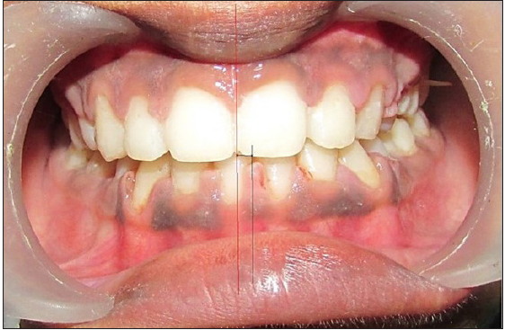
- Analysis of Joint Photographic Experts Group (JPEG) image using Digimizer software. Red line denotes maxillary dentition midline and black line denotes mandibular dentition midline.
RESULTS
The midline deviation was assessed using the Student’s t-test. The mean midline deviation in males was determined to be 1.12 mm, while in females, it measured 0.92 mm. The midline deviation was observed to be greater in males compared to females, and this difference proved to be statistically significant with a p-value of 0.034 [Table 1 and Figure 2].
| Mean Midline Deviation | S.D. | Min. | Max. | |
|---|---|---|---|---|
| Male | 1.12 | 0.75 | 0.03 | 3.70 |
| Female | 0.92 | 0.58 | 0.09 | 3.80 |
| t-test | 2.131 | |||
| p-value | 0.034 | |||
S.D.: Standard Deviation, Min: Minimum, Max: Maximum.
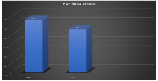
- Mean midline deviation among the study population.
DISCUSSION
The central focal point of an esthetically pleasing smile lies in the midline. A misalignment in the midline is easily noticeable to the patient. According to Boucher’s recommendation,[7] the central incisors’ long axis should align parallel to the face’s long axis, and the midline of the dental arch should ideally be positioned near the center of the face. Thilander et al. (2012)[8] conducted a study involving children at various developmental stages, revealing a 13.2% midline deviation in their sample. Meanwhile, Jayalakshmi et al. (2013)[1] noted a maxillo-mandibular dental midline discrepancy in nearly 80% of Indian students. In contrast, Anistoroaei et al. (2018)[9] reported a 20.70% midline deviation in orthodontic patients, and Hamid et al. (2020)[10] found a slightly higher percentage at 49.41%, particularly more pronounced in the maxilla than the mandible. In this comprehensive study, the mean distances between upper and lower dental midlines were determined to be 1.12 ± 0.85 mm in males and 0.92 ± 0.58 mm in females. Notably, in almost 80% of the population, the upper and lower dental midlines did not align. The standard deviation in our study was observed to be higher among males than females,and this difference proved to be statistically significant within the range of 0.03–3.80 mm. Contrary to the present study, Anistoroaei et al. (2018)[9] and Hamid et al. (2020)[10] found that the deviation of the dental midline was more frequent in females, though the difference was not statistically significant (p = 0.684). The studies by Sharma V[11] and Khan MF[12] recorded maxillary and mandibular midline deviations, as well as a substantial majority of the studies[13–15] exhibited a deviation between upper and lower dental midlines as well as anterior teeth midline, falling within the range of 0.03–3.80 mm.
CONCLUSION
A significant portion of the school-going school-going children in the North Bihar population exhibited deviations between the upper and lower midlines and the midline of anterior teeth, falling within the range of 0.03–3.80 mm. The prevalence of these deviations was higher in males than in females, and this difference proved to be statistically significant.
Ethical approval
The research/study complied with the Helsinki Declaration of 1964.
Declaration of patient consent
The authors certify that they have obtained all appropriate patient consent.
Financial support and sponsorship
Nil.
Conflicts of interest
There are no conflicts of interest.
Use of artificial intelligence (AI)-assisted technology for manuscript preparation
The authors confirm that there was no use of artificial intelligence (AI)-assisted technology for assisting in the writing or editing of the manuscript and no images were manipulated using AI.
REFERENCES
- Acceptable deviation between facial and dental midlines in dentate population. J Indian Prosthodont Soc. 2013;13:473-47.
- [CrossRef] [PubMed] [Google Scholar]
- Dental esthetics and the golden proportion. J Prosthet Dent. 1978;40:244-52.
- [CrossRef] [PubMed] [Google Scholar]
- Dental occlusion: A critical reflection on past, present and future concepts. J Oral Rehabil. 2008;35:446-45.
- [CrossRef] [PubMed] [Google Scholar]
- Digital dental photography. Part 1: An overview. Br Dent J. 2009;206:403-7.
- [CrossRef] [PubMed] [Google Scholar]
- Application of digimizer image analysis software in facial anthropometry. Iran J Ergon. 2020;8:61-71.
- [Google Scholar]
- Prosthodontic treatment for edentulous patients. IV. St. Louis: C.V. Mosby Co; 1975. p. :387-8.
- Posterior crossbite and temporomandibular disorders (TMDs): Need for orthodontic treatment? Eur J Orthod. 2012;34:667-73.
- [CrossRef] [PubMed] [Google Scholar]
- The prevalence of dental midline deviation in a group of orthodontic patients. Rom J Oral Rehabil. 2018;10:176-82.
- [Google Scholar]
- Prevalence of dental midline shifting in a sample of orthodontic patients in Erbil city. Journal of University of Raparin. 2020;7:545-55.
- [Google Scholar]
- Correlation of facial to dental midline and maxillary to mandibular midline in Karnataka population. Ann Essen Dent. 2010;3:20-4.
- [Google Scholar]
- Frequency of dentofacial asymmetries: A cross-sectional study on orthodontic patients. J Ayub Med Coll Abbottabad. 2014;26:129.
- [PubMed] [Google Scholar]
- Prevalence of midline shift in orthodontic patients. International Journal of Current Innovation Research. 2015;1:287-90.
- [Google Scholar]
- Corelation of facial and dental midlines in dentate population of North Bihar. Jr of Population Therapeutics & Clinical Pharmacology. 2023;30:472-7.
- [Google Scholar]


