

Translate this page into:
AN IMPACTED SUPERNUMARY TOOTH: A STARTLING PRESENTATION
Corresponding Author : Dr. Smita Lahane
This article was originally published by Indira Gandhi Institute of Medical Science and was migrated to Scientific Scholar after the change of Publisher.
Abstract
Supernumerary dentition has been found to occur in various areas of the head and neck. Though they are frequently found along the dental arch. This case report details an impacted ectopic supernumerary molar tooth. Here, we present an unusual, accidental case of impacted ectopic supernumerary tooth at angle of mandible of 60-year-old male. Radiographically, OPG and CT scan revealed a benign appearing tooth like structure at the angle of mandible on right side.
Keywords
Ectopic Tooth
Supernumerary Tooth
OPG
CT Scan.

INTRODUCTION:
Tooth eruption is a process in which the forming tooth migrates from its intraosseous location in the jaw to its functional position within the oral cavity. Various eruption problems arise during the transitional period of tooth eruption and ectopic eruption is one of them.1 In ectopic eruption the tooth does not follow its usual route. The entity is been occasionally discovered in unusual orientations or at a distance from their normal anatomic position. The etiology of ectopic eruption can be due to disturbance of the differential growth pattern of the individual. Different tissues and organs grow at different rates and at different times. A delicate balance normally exists between the timing and rate of growth. Differential growth is the origin for normal and harmonious completion of various physiologic processes including the eruption of teeth. Whenever this balance is distressed, whether due to congenital factors or environmental interferences, an abnormal situation develops. It is, therefore, obvious that whatsoever the etiological factor, a disturbance in the balance between the rate of jaw growth, the rate of eruption of the first molars, and/or sizes of the teeth produces ectopic eruption16. Ectopic and supernumerary teeth have been rarely described in non-dental and non-oral sites8 such as in mesial, distal apically, palatally from the normal position and rarely in the orbit, maxillary antrum, nasal cavity, nasal septum, mandibular condyle, coronoid process, palate, and chin.2 Mandibular molars are commonly involved, specially third molars, which are impacted far away from their original sites. In most cases, conditions such as dentigerous cysts or nasal and maxillofacial symptoms may associated with ectopic teeth. In the present study, we report a case of an ectopic mandibular molar located at the angle of mandible with OPG and Cone Beam Computed Tomography (CBCT) that seems to have been displaced by neither cyst nor tumor and not associated with any pathology3.
CASE REPORT:
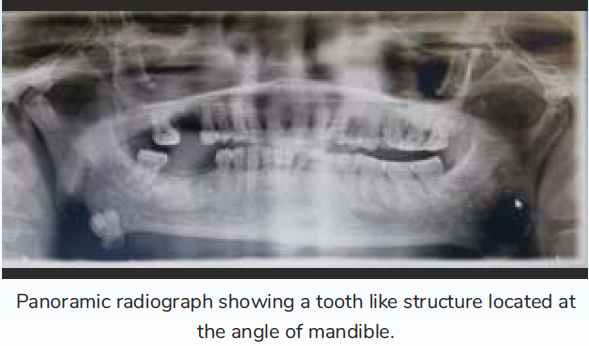
A 60-year-old male patient visited the Department, complaining of pain in the right upper and lower back teeth region. After intraoral examination, he diagnosed as trigeminal neuralgia involving right side of face maxillary and mandibular division after Diagnostic block testing with 2% of plain xylocaine and prescribed Tab Carbamazepine 200 mg twice daily and on follow-up visits showed improvement in the complaints. The panoramic radiograph of the patient shown an impacted and displaced tooth at the right angle of mandible. Tooth was identified as ectopic, as third molar was on position and other two molars were existed on the right side. Patient has no signs & symptoms for the same.
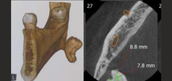
Further was investigated with Cone Beam Computed Tomography (CBCT) that has shown tooth morphology like structure at the angle of mandible of size approx. 18× 8.5 mm lingually 6.3 mm away from mandible suggestive of ectopic distomolar.
DISCUSSION:
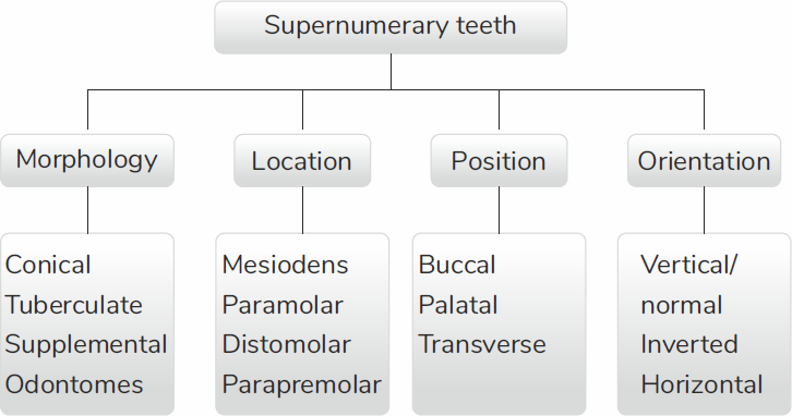
Supernumerary tooth or an addition to the regular number of teeth is a rare developmental variance that can occur in any area of the dental arch. This occurrence is also known as hyperdoncia and can occur in solitary or multiple form, may be unilateral or bilateral, and affect one or both jaws and have a remarkable predilection for maxilla over mandible. These teeth are more dominant among men than women in a proportion of 2:1.5 the frequency of supernumerary teeth in deciduous dentition is 0.3% to 0.8% and 1.5% to 3.5% in the permanent dentition4. They are most commonly located in the maxilla, the anterior medial region, where 80% of all supernumerary teeth are found. More infrequently, they can be located in the inferior premolar zone, superior distomolar zone, superior premolar, superior canine zone, inferior distomolar, and inferior incisor6. Numerous supernumerary teeth are frequently present when a syndrome is involved. Yusof et al suggested that it may be occasional to find multiple supernumerary teeth without an associated syndrome. Communal syndromes showing multiple supernumerary teeth along with other conditions include Gardiner's syndrome, cleft lip and palate and cleidocranial dysostosis12. The meticulous etiology of supernumerary teeth is unknown, however, numerous theories have been hypothesized to try to explain their presence: the phylogenetic theory as a regression to the anthropoids whose dental formula had more teeth, an abnormal reaction to a local traumatic episode, the autonomic recessive inheritance or linked to the X chromosome, environmental factors, dichotomy of the tooth germ and the theory of hyperactivity of the dental lamina, are the most accepted10. Supernumery teeth classified based on morphology (conical, tuberculate, supplemental, and Odontomes), location (Mesiodens, Paramolar, distomolar, and Parapremolar), position (buccal, palatal, and transverse), orientation (vertical or normal, inverted, transverse, or horizontal)11.
The presence of supernumerary teeth is a common dental anomaly, but the occurrence of Paramolars and Parapremolar is relatively uncommon. Supernumerary teeth can be asymptomatic and are diagnosed as an incidental finding during radiographic examination. On the other hand, Ectopic supernumery tooth may be asymptomatic or existing with a variety of signs and symptoms, such as rhinorrhea, nasal obstruction, nasal congestion, pain, nasal bleeding, chronic inflammation, persistent discharge and crusting, septal abscess and fistula formation, and external nasal deviation. These symptoms can be established, recurrent, and impassive to systemic therapy with antibiotics and corticosteroids9. In present study we advise follow up for the ectopic distomolar at the angle of mandible. The frequency of Mesiodens (47 - 67%), premolar (8-9%), distomolar (26%), Paramolars (15%), lateral incisor (2.05%), and canine (0.40%). 6,7
Effects of supernumerary teeth on the developing dentition differ. There may be no effect of supernumery tooth or they discovered accidentally on radiograph. Crowding may be apparent due to an increased number of erupted teeth. Failure of eruption of adjacent permanent teeth is the most recurrent occurrence and occurs in 30 to 60 per cent of cases. The supernumerary or adjacent teeth may be displaced and ectopic eruption of either is not uncommon12. Dentigerous cysts are the most common jaw lesion in this category(65%) mostly impacted third molar, followed by calcifying epithelial odontogenic tumors (52% - 60%) predominantly impacted third molar and calcifying odontogenic cysts (10% - 20%) predominantly impacted canine, The maximum incidence of association with an impacted tooth was seen for unicystic ameloblastomas (50% - 80%)predominantly impacted third molar, adenomatoid.
Odontogenic tumors (73%) predominantly impacted canine, odontomas, and dentigerous cysts (65%)15. The association of a dentigerous cyst with supernumerary teeth constitutes only 5-6% of all dentigerous cysts12. The cysts related with these teeth can persist for years without symptoms and may be detected only by routine imaging8. In other cases, patients become symptomatic with signs of sinus disease such as swelling, facial pain, nasolacrimal duct obstruction and headache. Moreover, a large maxillary cyst can cause orbital and sinonasal symptoms13.
Conclusion:
The most frequently impacted supernumerary teeth are maxillary canines and mesiodens. They are more common in males than females. In our case the site of impacted supernumerary tooth is unusual which is at the angle of mandible on right side and it is not associated with any pathology and was asymptomatic. It is rare to ensue and careful investigation needed to rule out this findings.
REFERENCES:
- Ectopic eruption - A review and case report Contemporary Clinical Dentistry. 2011;2(1):3-7.
- [CrossRef] [PubMed]
- Ectopic Premolar Tooth in the Sigmoid Notch. 2016:3. Article ID 6426523
- [CrossRef] [PubMed] [Google Scholar]
- Process of ectopic tooth formation in the maxillary sinus: follow-up observation of one case journal of International Medical Research. . 2019;47(12):6356-6364.
- [CrossRef] [PubMed] [Google Scholar]
- Ectopic Supernumerary Tooth at the Anterior Nasal Spine- A Developmental Glitch Journal of Clinical and Diagnostic Research. . 2015;9(11):ZJ01-ZJ02.
- [Google Scholar]
- Prevalence, etiology, diagnosis, treatment and complications of supernumerary teeth. J Clin Exp Dent. 20142014;6(4):e414-e418.
- [CrossRef] [PubMed] [Google Scholar]
- Paramolar-A supernumerary molar: A case report and an overview. Dent. Res. J. 2012;9:797-803.
- [Google Scholar]
- Retrospective study of 145 supernumerary teeth. Med Oral Patol Oral Cir Bucal. 2006;11:E339-4.
- [Google Scholar]
- Supernumerary, ectopic tooth in the maxillary antrum presenting with recurrent haemoptysis. 2010. Head Face Med. 26:6.
- [CrossRef] [PubMed] [Google Scholar]
- Gurenlian, Managing Supernumerary and Ectopic Teeth. J multidisciplinary care. 2021;11
- [Google Scholar]
- Multiple hyperodontia: Report of a case with 17 supernumerary teeth with non syndromic association. Med Oral Patol Oral Cir Bucal. 2009;14(5):E229-31.
- [Google Scholar]
- Supernumerary Teeth: Review of the Literature with Recent Updates Hindawi Publishing Corporation Conference Papers in Science. 2014:6. Article ID 764050
- [CrossRef] [Google Scholar]
- Dentigerous cyst associated with an impacted anterior maxillary supernumerary tooth. BMJ Case Rep 2013
- [CrossRef] [PubMed] [Google Scholar]
- Odontogenic Cyst From an Ectopic Supernumerary Tooth Impacted in the Orbital Floor Journal of Advanced Oral Research. 2019;10(2):165-169.
- [CrossRef]
- Dentigerous cysts associated with impacted supernumerary teeth in the anterior maxilla. Exp Ther Med. 2011;2(5):805-809. Epub 2011 May 18
- [CrossRef] [PubMed] [PubMed Central] [Google Scholar]
- Jaw lesions associated with impacted tooth: A radiographic diagnostic guide. Imaging Sci Dent. 2016;46(3):147-157.
- [CrossRef] [PubMed] [Google Scholar]
- Ectopic eruption - A review and case report. Contemp Clin Dent. 2011;2(1)::3-7.
- [CrossRef] [PubMed] [Google Scholar]


