Translate this page into:
Morphological Characteristics of the Incisive Papilla in Pediatric Forensic Odontology: A Cross-Sectional Study

*Corresponding author: Dr. Santhosh Priya AKR, Department of Paediatric and Preventive Dentistry, Sathyabama Dental College and Hospital, Chennai, Tamil Nadu, India. santhosh.appiya@gmail.com
-
Received: ,
Accepted: ,
How to cite this article: AKR SP, James B, C VR, Saleem S, K SSK, PL S. Morphological Characteristics of the Incisive Papilla in Pediatric Forensic Odontology: A Cross-Sectional Study. Dent J Indira Gandhi Int Med Sci. 2025;4:6-9. doi: 10.25259/DJIGIMS_35_2024
Abstract
Objectives
Forensic odontology plays a critical role in identification of individuals through dental evidence. The study aims to evaluate the prevalence and morphological characteristics of incisive papilla among children aged 3-6 years and assess its potential as a reliable tool for racial identification.
Material and Methods
A cross-sectional study was conducted involving 462 pediatric subjects recruited from Sathyabama Dental College and Hospital. Participants were assessed for incisive papilla morphology using Ortman and Tsao’s classification.
Results
The study sample comprised 262 males (56.7%) and 199 females (43.3%). The most common shape of the incisive papilla was pear (51.5%), followed by rectangular (34.1%). No statistically significant differences in papilla shape were observed between genders (p = 0.103).
Conclusion
The findings indicate that the pear shape is the predominant morphology of the incisive papilla in the Chennai population, and its shape can serve as a reliable forensic parameter for identification in dentulous individuals without surgical alterations. Further research is warranted to enhance understanding of its applicability in forensic odontology.
Keywords
Incisive papilla
Forensic odontology
Racial identification
INTRODUCTION
Forensic odontology represents a specialized domain within dentistry that focuses on the meticulous management and analysis of dental evidence, along with the comprehensive assessment and presentation of dental findings to uphold the principles of justice. The dental structures are recognized as some of the most resilient and well-guarded components of the human body, capable of withstanding decomposition and elevated temperatures, thereby ranking among the final anatomical structures to undergo deterioration post-mortem.[1] The foundational principle underpinning dental identification resides in the premise that no two oral cavities exhibit identical characteristics, with teeth possessing distinctive features that are unique to each individual. In pediatric populations, the incisive papilla emerges as a pivotal landmark for forensic identification, given that its position and morphological attributes can yield critical insights into individual dental characteristics.
Moreover, beyond its significance in forensic identification, the morphological traits of the incisive papilla can facilitate a deeper understanding of ethnic variations in dental anatomy. The incisive papilla is defined as a fleshy protrusion of the palatal mucosa situated posterior to the maxillary central incisors. Typically, it manifests with a hue that is markedly intense in redness when juxtaposed with the surrounding pale pink palatal mucosa. This anatomical structure maintains a substantial relationship with the contact points of the teeth, the interdental spaces, and the proximal surfaces of the cementoenamel junction. The incisive papilla is distinguished by its uniqueness, with its internal positioning, regional variations, and resilience to antemortem alterations, rendering it a potentially reliable source for identification objectives. Its anatomical placement within the oral cavity provides a degree of protection against both trauma and extreme thermal exposure. The configuration of the papilla remains stable even subsequent to orthodontic interventions.[2] Very limited literature exists regarding the influence of incisive papilla in forensic dentistry. Consequently, this investigation aspires to determine the prevalence of the morphological characteristics of the incisive papilla within the Chennai population. Additionally, it endeavors to examine the potential of the shape of the incisive papilla as a trustworthy forensic instrument for racial identification within the Chennai demographic.
MATERIAL AND METHODS
The research cohort consisted of 462 pediatric subjects aged between 3 and 6 years, recruited from Sathyabama Dental College and Hospital.
Before engaging in the study, all participants were presented with a consent form, and measures were taken to ensure their confidentiality by keeping their personal information anonymous. Healthy children between 3 and 6 years with no dental anomalies were included in the study.
The subjects were positioned comfortably in a dental chair, and images of the incisive papilla were captured using a camera, a dental mirror, and adequate illumination. A trained dentist evaluated the morphology of the incisive papilla. The categorization of the incisive papilla shape was performed in accordance with Ortman and Tsao’s classification system.[3]
Statistical analysis was executed employing SPSS IBM software version 25. Descriptive statistics were utilized to ascertain the frequency distribution and corresponding percentages. The Chi-square test was employed to investigate the association between the shape of the incisive papilla and the gender of the study participants. A p-value <0.05* was deemed statistically significant.
RESULTS
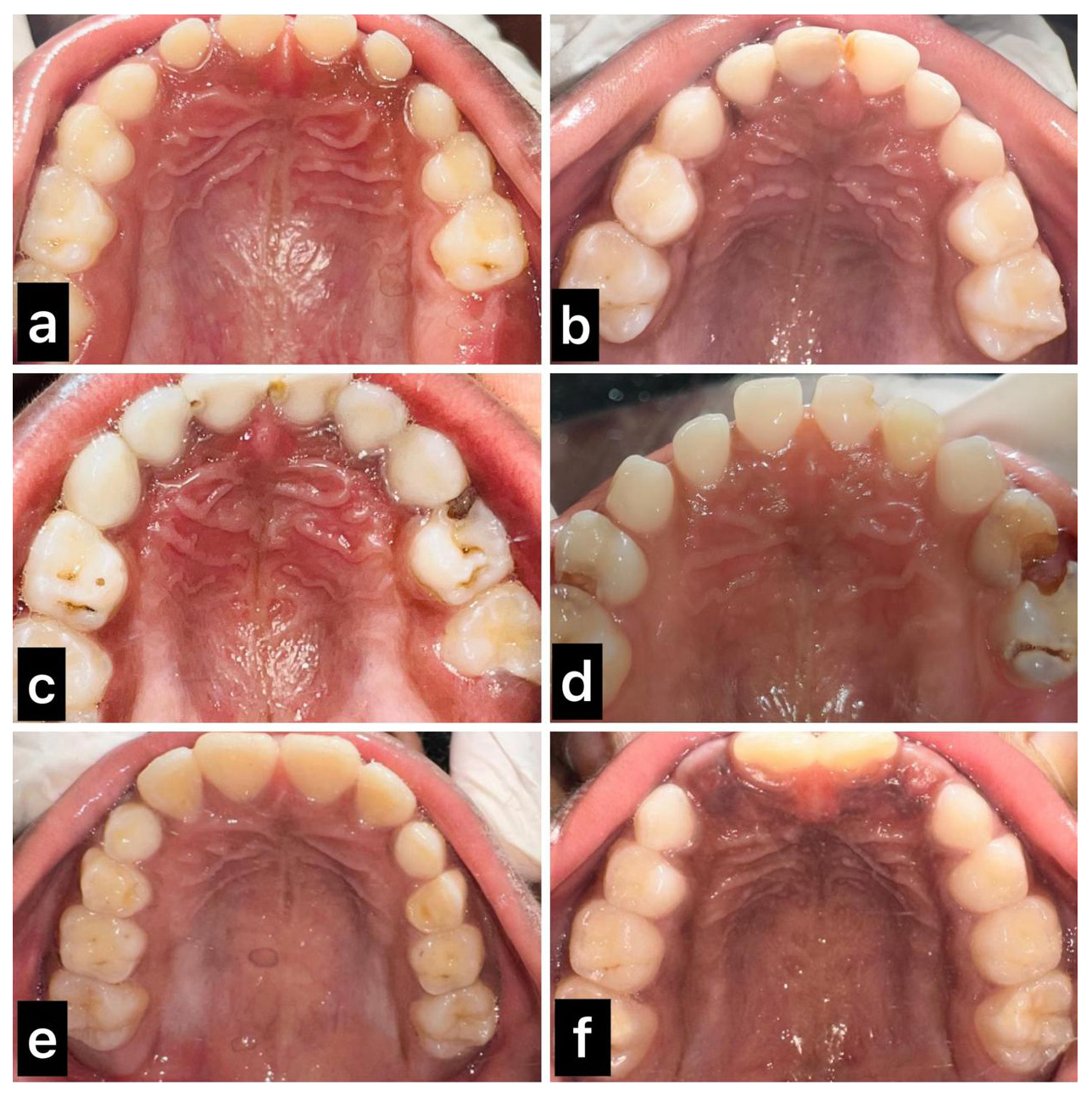
This cross-sectional study was conducted on a study sample of 462 participants. Among the total 462 study participants, the majority of them were males, representing 262 (56.7%), and 199 (43.3%) were males. The shape of the incisive papilla was evaluated, depending on the shape – rectangular, pear, oval, irregular, triangular, inverted pear, or no papilla [Figure 1]. Our study shows that a pear shape was the most common, followed by a rectangular shape. The irregular shape was not recorded in any children. The study population was also analyzed based on the shape of the incisive papilla and gender, which was statistically not significant [Table 1].

- (a) Rectangular, (b) triangular, (c) oval, (d) inverted pear, (e) no papilla, (f) pear.
| Incisive papilla shape | Female n (%) | Male n (%) | P value |
|---|---|---|---|
| Inverted pear | 0 (0%) | 4 (1.5%) | 0.103 |
| Rectangular | 69 (34.5%) | 88 (33.6%) | |
| No papilla | 4 (2%) | 0 (0%) | |
| Oval | 14 (7%) | 15 (5.7%) | |
| Pear | 102 (51%) | 136 (51.9%) | |
| Triangular | 11 (5.5%) | 19 (7.3%) | |
| Irregular | 0 (0%) | 0 (0%) |
DISCUSSION
In the field of forensic dentistry, the anatomical composition and spatial orientation of the incisive papilla are acknowledged as critical factors for the identification of individuals through dental remains. The anatomical characteristics of the incisive papilla have been classified by various authors, including the classifications proposed by Soloman and Arunachalam, as well as by Ortsman and Tao, among others.[3-5] In the current investigation, we have opted to employ the Ortsman and Tao classification due to its simplicity and reliability. Our findings revealed that the pear shape, followed by the rectangular shape, predominates among the Chennai population. This observation aligns with the studies conducted by Soloman et al.[4] and Mustafa et al.,[6] which also focused on South Indian populations. However, our findings stand in contrast to the research conducted by Samarth Kumar et al., which posited that the cylindrical shape is prevalent within the Moradabad (Uttar Pradesh) population.[7] As children undergo continuous growth, variations in orodental morphology are observed, particularly during the transition from deciduous to permanent dentition; nevertheless, the shape of the papilla remains unchanged. Thus, the configuration of the papilla may serve as a valuable criterion for racial determination. This suggests that while certain morphological traits may vary across different regions, the papilla’s shape could provide a consistent marker for identifying racial characteristics in dental studies.
The distribution of the study population was further analyzed in relation to the shape of the papilla and gender. The results indicated that a majority of individuals across both genders exhibited pear-shaped papillae, revealing no statistical significance, akin to the findings reported by Krisnanand et al.[1] This outcome contrasts with the work of Mustafa et al.,[6] who identified a gender disparity, which may be attributed to the fact that their study was conducted within a Jordanian population. Further investigation into the environmental factors influencing these morphological traits could shed light on the observed differences and enhance our understanding of dental anthropology.
Upon reviewing the existing literature, it is evident that orthodontic interventions do not modify the shape of the papilla. However, in instances where anterior teeth are extracted or in edentulous patients, the papilla tends to adopt a rounded configuration and migrates anteriorly, resulting in alterations to its original morphology.[4] In cases involving surgical interventions, such as the removal of a nasopalatine cyst, the incisive papilla may undergo collapse. Consequently, in such scenarios, the papilla cannot be regarded as a reliable source of information within the domain of forensic dentistry. This highlights the importance of considering individual patient histories and treatment outcomes when utilizing the papilla as a reference point in forensic analyses.[8]
Therefore, the anatomical characteristics of the incisive papilla play a pivotal role in forensic dentistry, particularly in the identification of individuals through dental remains. Our study, which utilized the Ortsman and Tao classification, identified a predominance of pear-shaped and rectangular papillae within the Chennai population, corroborating previous research while also highlighting regional differences. The consistency of the papilla’s shape, despite variations in orodental morphology during growth, suggests its potential utility as a marker for racial determination in dental studies. Furthermore, the lack of significant gender disparity in our findings contrasts with some literature, indicating a need for further exploration into the environmental and genetic factors influencing these morphological traits. The review of existing literature underscores the stability of the papilla’s shape in the absence of orthodontic interventions while also noting the impact of surgical procedures on its morphology. Therefore, while the incisive papilla can serve as a valuable tool in forensic analyses, careful consideration of individual patient histories and treatment outcomes is essential to ensure its reliability as a forensic marker. Continued research in this domain will enhance our understanding of dental anthropology and improve the application of these findings in forensic contexts. Moreover, interdisciplinary collaboration between forensic scientists and dental professionals will be crucial in refining methodologies and establishing standardized protocols for the assessment of the incisive papilla’s characteristics.[9,10]
Limitation of the study
The study’s sample size could be increased for more robust prevalence rates. The gender distribution was imbalanced, with more males than females, affecting the representation of both sexes in the findings.
CONCLUSION
-
1.
Prevalence of shapes: In the Chennai population, the pear shape of the incisive papilla was the most commonly observed, followed by the rectangular shape.
-
2.
Gender analysis: No statistically significant differences in incisive papilla morphology were found based on gender.
-
3.
Forensic relevance: The shape of the incisive papilla can be considered a reliable forensic parameter for identifying dentulous individuals without prior surgical alterations.
Implications for future research: Findings suggest a need for further studies to explore the morphological characteristics of the incisive papilla in diverse populations and their potential applications in forensic odontology.
Ethical approval
The research/study was approved by the Institutional Review Board with approval number: 325/IRB-IBSEC/SIST.
Declaration of patient consent
The authors certify that they have obtained all appropriate patient consent.
Financial support and sponsorship
Nil
Conflicts of interest
There are no conflicts of interest.
Use of artificial intelligence (AI)-assisted technology for manuscript preparation
The authors confirm that they have used artificial intelligence (AI)–assisted technology for assisting in the writing or editing of the manuscript or image creations.
REFERENCES
- Establishing the reliability of incisive papilla and palatal rugae patterns in individual identification. J Oral Maxillofac Pathol. 2023;27:779.
- [CrossRef] [PubMed] [PubMed Central] [Google Scholar]
- Characteristic changes of the palatal rugae following orthodontic treatment. Egypt J Forensic Sci. 2023;13:14. Available from: https://doi.org/10.1186/s41935-023-00334-5
- [CrossRef] [Google Scholar]
- Relationship of the incisive papilla to the maxillary central incisors. J Prosthetic Dentistry. 1979;42:492-6.
- [CrossRef] [Google Scholar]
- The incisive papilla: A significant landmark in prosthodontics. J Indian Prosthodont Soc. 2012;12:236-47.
- [CrossRef] [PubMed] [Google Scholar]
- A study of the rugae pattern and the shape of the incisive papilla in Sri Lankan population. Ceyl Dent J. 1978;9:11-21.
- [Google Scholar]
- Morphometric study of the hard palate and its relevance to dental and forensic sciences. Int J Dent. 2019;2019:1687345.
- [CrossRef] [PubMed] [PubMed Central] [Google Scholar]
- Evaluation of papillo-incisal distance in different arch forms and with different shapes of incisive papilla in Moradabad population - A descriptive study. J Dent Probl Solut. 2021;8:42-6.
- [CrossRef] [Google Scholar]
- The Incisive Papilla: The Basis of a Technic to Reproduce the Positions of Key Teeth in Prosthodontia. Journal of Dental Research. 1948;27:661-8.
- [Google Scholar]
- Correlation between the size of the incisive papilla and the distance from the incisive papilla to the maxillary anterior teeth. J Dent Sci. 2016;11:141-5.
- [CrossRef] [PubMed] [PubMed Central] [Google Scholar]
- Assessment of The Relationship of Incisive Papilla to Maxillary Central Incisor and Canine-Papilla-Canine Line among the Dentate population of Central Nepal. Kathmandu Univ Med J (KUMJ). 2017;17:150-4.
- [PubMed] [Google Scholar]


